We work with eye doctors and other doctors such as primary care physicians to assist in providing the best patient care available. We are committed to helping you grow your practice.
Figure 1. Ray tracing simulation for the automated eye model.
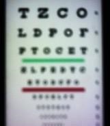
Figure 2. Images from the automated eye model. (a) image requiring correction. (b) corrected image.
Abstract
Purpose : An automated human eye model was designed and built for test and validation of refraction measurement equipment. It further enables visualization of various ocular conditions including, but not limited to, myopia, hyperopia, and accommodation. The automated eye model can serve as an educational tool at ophthalmology and optometry schools as well as a demonstration tool in clinics and offices.
Methods : The automated eye model is comprised of an optical system that is aimed at providing refraction control and a camera serving as the retina. It is built based on the human eye anatomy. The optical system consists of a fixed lens and a Stokes pair serving as a cornea equivalent, an iris that controls the pupil size of the automated eye model, and a variable focus lens serving as the eye lens. A camera is mounted on a linear stage to account for different intra-ocular distances associated with various conditions. The refraction control is achieved by changing the variable lens power or changing the intra-ocular distance for the spherical part and controlling the rotational orientation of the Stokes pair for the cylinder and axis parts.
Results : Figure 1. presents a ray tracing analysis of the automated eye model. The three colors represent different field angles corresponding to the human macular field of view of ±9°. The optical train described in the Methods section is simulated. The fact that the image plane is flat (as is a camera) and not curved presents a field dependent aberration. In future designs an aspheric lens could be used to mitigate this effect. Figure 2.a. presents an image of a visual acuity chart as seen through the automated eye model for an example refraction of -3.25D, figure 2.b. presents the same image as seen with the same automated eye model settings with the appropriate corrective lens.
Conclusions : The automated eye model is a tool that enables the simulation of the human eye with respect to refractive functionality. It allows for independent control of the spherical, cylindrical and axis values enabling observation of effects on visual outcome with the application of various corrective lenses. The device also allows for validation of refraction equipment accuracy. Future work will address aspects related to optical quality, scale, cost and further functionality.
This abstract was presented at the 2019 ARVO Annual Meeting, held in Vancouver, Canada, April 28 – May 2, 2019.